


Submit a comment
Using chitosan from shrimp shells and alginate in seaweed, Master Vu Thanh Binh created an artificial skeleton to restore missing bones.
MSc. Binh (34 years old), currently works at the Laboratory of Tissue Engineering and Regenerative Medicine, Faculty of Biomedical Engineering, International University (Ho Chi Minh City National University). He and his research team have succeeded in creating a scaffold that helps bone cells adhere, proliferate and calcify to restore defective bone.
Dr. Binh said that the body's natural bone tissue has polymer components, which are collagen fiber bundles and hydroxyapatite (calcium phosphate). These components create a bone tissue structure that can withstand loads, perform support functions, and create a marrow cavity... Based on this, the group created natural polymers using chitosan found in shrimp and crab shells along with alginate found in seaweed.
Chitosan and alginate are combined with a polymer found in synovial fluid called hyaluronic acid, which helps increase elasticity and reduce damage to the joint ends. "These three materials need to be linked together to form an artificial bone scaffold," said Master Binh. The bone scaffold created in the laboratory has a structure that is closest to natural bone tissue.
According to the group, the bonding of raw materials can use additives. However, this method has the disadvantage of residual exogenous cross-linking substances, which can be toxic to bone cells. Therefore, the research group implemented a method of changing the structure of raw materials by adding functional groups, giving them the ability to self-bond without the use of additives.
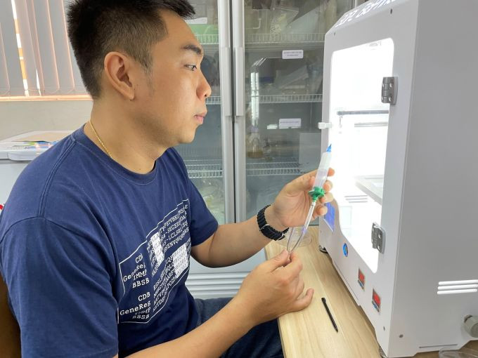
Master Vu Thanh Binh tests a 3D biological printer that uses gel as ink to create artificial skeletons.
From the gel-like material, the solidified skeleton creates a porous structure that helps natural bone tissue cells to adhere to the skeleton to proliferate. This artificial skeleton is biodegradable (disappears after human bone cells adhere and grow). This bone cell can secrete a matrix to create its own bone skeleton to fill the missing bone. Based on the properties and location of the missing bone tissue, the team can adjust the biodegradation time to match the time bone cells secrete and create the skeleton.
According to Dr. Binh, depending on the type of bone and the volume of bone loss, bone cells need several months to years to regenerate a new frame and completely restore the missing area.
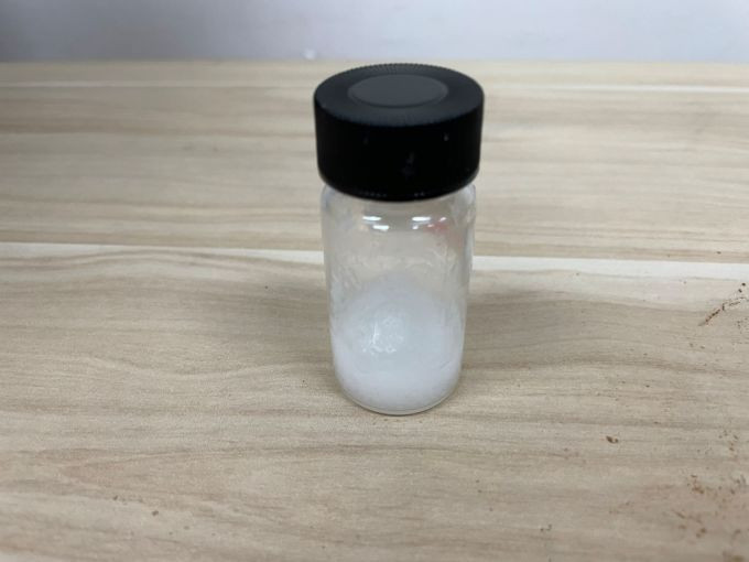
Artificial bone material of natural origin exists in gel form developed by research team
The team tested the technique on mice by anesthetizing them, drilling a hole in the skull cap to create a bone defect without damaging their brains. Then, they injected gel into the area where the mice had bone defects. When the mice woke up, their vital signs such as weight, diet, movement, etc. were monitored for a month.
The author said that because the material is in gel form, when it enters the skull cap, it can fill the defect regardless of its shape. In a short time, the gel will solidify and begin the process of creating an artificial skeleton. After one month, the team humanely euthanized the mouse, performed brain surgery to evaluate the ability to regenerate bone tissue based on the artificial skeleton using biological tissue staining methods. The results showed an 80-90% filling rate of the defect on the mouse skull cap, with high biocompatibility.
The results are the basis for the group to be able to test on large animals with pathological conditions more similar to humans, strengthening the scientific evidence for application on humans. However, according to Master Binh, from animal models to human applications is quite a distance, going through many processes, procedures and Ethics Councils to prove the effectiveness and safety of the product.
In the immediate future, the research team wants to develop the scaffold gel into a commercially viable bioink. The bioprinted gel can form artificial scaffolds for biomedical research and animal testing, replacing expensive imported products.
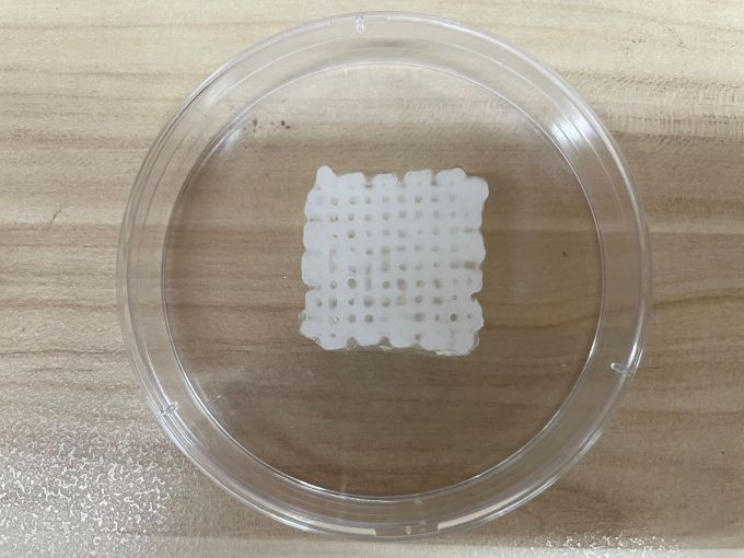
The shape of the artificial bone after being printed from the 3D biological printer
According to Associate Professor, Dr. Nguyen Thi Hiep, Head of the Department of Biomedical Engineering, International University (Ho Chi Minh City National University), the research direction of artificial bones from natural materials has been carried out by scientists around the world for over 10 years. In Vietnam, there have been a number of studies carried out at institutes and schools, aiming at personalized treatment of bone lesions in humans. This overcomes the problem of treatment from available bone frames, which in many cases are not suitable for each person. The group created a gel that, after being injected into the bone defect area, can be shaped and suitable for anyone.
However, Associate Professor Hiep said that the research needs to conduct testing steps on large animals to assess safety and feasibility on larger bone sizes, moving towards clinical trials on humans. "With this research, we are participating in seeking funding sources to conduct larger-scale testing steps, perfecting technological solutions, and contributing to the field of precision medicine in the country," Associate Professor Hiep said.
According to VnExpress